Dr. Jinal Gore
What is hypertension?
Blood pressure is the force exerted by circulating blood against the walls of the body’s arteries, the major blood vessels in the body. Hypertension is when blood pressure is too high. Hypertension significantly increases the risks of heart, brain, kidney and other diseases.
Kearney and colleagues estimated that the prevalence of hypertension in 2000 was 26% of the adult population globally and that in 2025 the prevalence would increase by 24% in developed countries and 80% in developing countries.
What is hypertensive retinopathy?
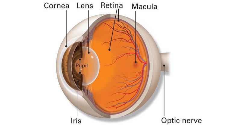
The retina is the tissue layer located in the back of your eye. It transforms light into nerve signals that are then sent to the brain for interpretation.A high blood pressure for a long duration results in narrowing of blood vessel lumen resulting in damage to retina and optic nerve due to less blood supply. This condition is called hypertensive retinopathy (HR).
What are the symptoms of hypertensive retinopathy?
It’s called a “silent killer” because it usually has no symptoms. In late stages you may have reduced vision.
What are the conditions associated with increased risk of hypertensive retinopathy?
Prolonged high blood pressure, presence of hypertension in family members, heart disease, atherosclerosis, diabetes, smoking, high cholesterol, obesity, lack of physical activity, consuming excessive salt in diet, stressful lifestyle and excess alcohol consumption.
How is hypertensive retinopathy diagnosed?
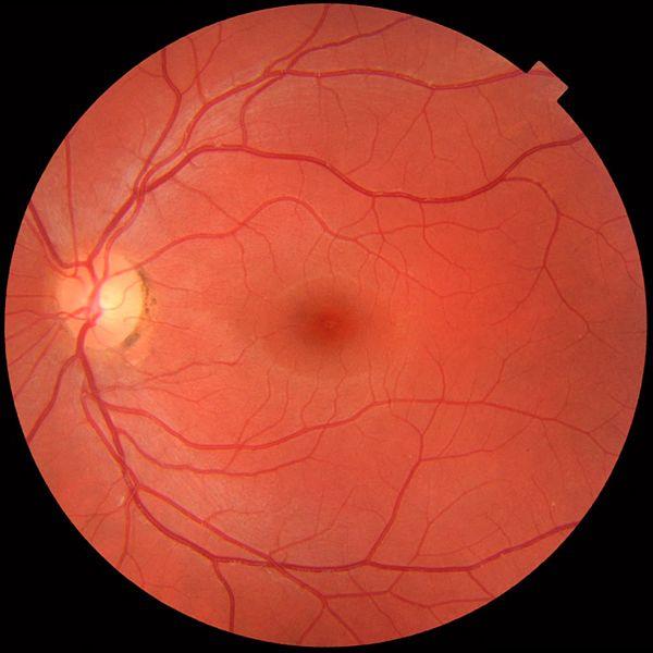
Your doctor will use a tool called an ophthalmoscope mounted on head to examine your retina. This instrument shines a light through your pupil to examine the back of your eye for signs of narrowing blood vessels or to see if any fluid is leaking from your blood vessels. This procedure is short and painless.
Other tests for hypertensive retinopathy are:
- Optical coherency tomography (OCT)
This test helps to obtain cross sectional scans your retina to look for fluid accumulation in it which may be the cause of reduced vision. This is a painless procedure and the scans are obtained with the help of OCT machine while patient is in sitting position.
- Fluorescein angiography
As the name suggests, this test helps in seeing pattern of blood flow through retina. In this procedure, your doctor will apply special eye drops to dilate your pupils, fluorescein dye is injected in your blood through a cannula and multiple pictures of your retina are taken as the dye moves through circulation.
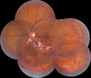
Grading of hypertensive retinopathy
The extent and severity of hypertensive retinopathy is generally represented on a scale of 1 to 4. The scale is called the Keith-Wagener-Barker Classification System. On the lower end of the scale, you may not have any symptoms. At grade 4, however, your optic nerve may begin to swell and cause more serious vision problems. High-grade retinopathy tends to indicate serious blood pressure concerns.
Complications of hypertensive retinopathy
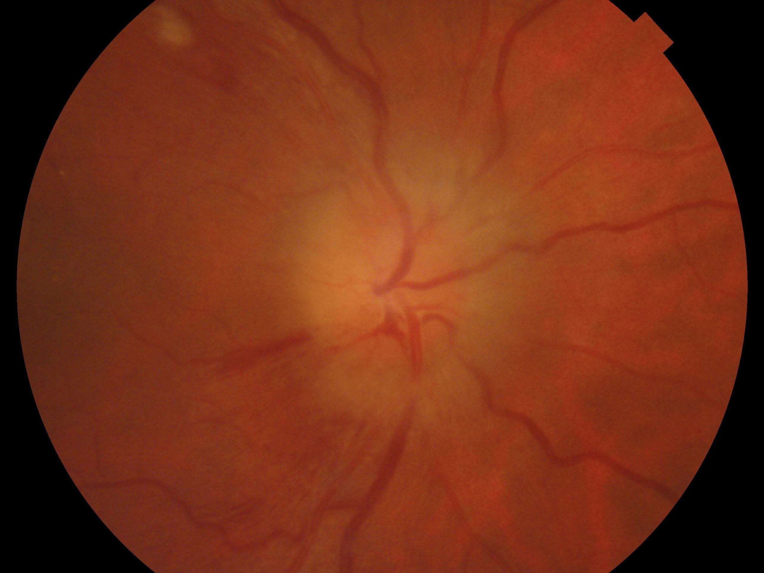
- Retinal artery occlusion: The central retinal artery supplies blood to inner retina. Blockage of this artery by blood clot results absence of oxygenated blood to reach the retinal tissues thus, causing sudden and severe loss of vision.
- Retinal vein occlusion: The retinal vein carries impure blood away from retina to the heart. Its blockage by any blood clots causes engorgement of impure blood in retina, fluid collection, ischemia which results in loss of vision.
- Ocular ischemic syndrome: In this condition there is narrowing of carotid artery which is the main artery supplying head resulting in subsequent damage to retina.
- Ischemic optic neuropathy: This condition occurs due to obstruction of blood vessels supplying the optic nerve as a result of high blood pressure, thereby damaging the optic nerve.


